Medical AI + XR Spatial IntelligenceDoctor-led explanation

Making Medical Imaging Spatially Understandable.

Pholoid turns medical imaging, AI imaging outputs, and physician knowledge into spatial systems for education, explanation, case discussion, and collaboration.

AI reads pixels. Pholoid helps people understand the result in space.

Problem

Medical imaging and AI results still live too flat.

CT, MRI, DICOM slices, segmentation masks, lesion boundaries, and physician notes are rich with spatial meaning. But flat reports and static screens make that meaning hard to teach, explain, review, and discuss.

Pholoid focuses first on practical, commercially legible workflows where spatial understanding improves education, patient communication, case collaboration, and AI result explanation.

2D screensStatic reportsFragmented discussion

DeepView XR Engine

The spatial engine beneath Pholoid.

DeepView XR Engine converts medical imaging data, AI segmentation outputs, and physician annotations into interactive spatial medical models for teaching, communication, and collaborative review.

DICOM importAI segmentationLayered anatomy2D / 3D linkageDoctor annotationMulti-device access

Product Matrix

One engine, four focused commercial paths.

Pholoid starts with high-value, lower-regulatory-pressure workflows: education, communication, discussion, collaboration, and partner explanation layers for medical AI and hospital software ecosystems.

Core imaging spatial engine

DeepView XR Engine

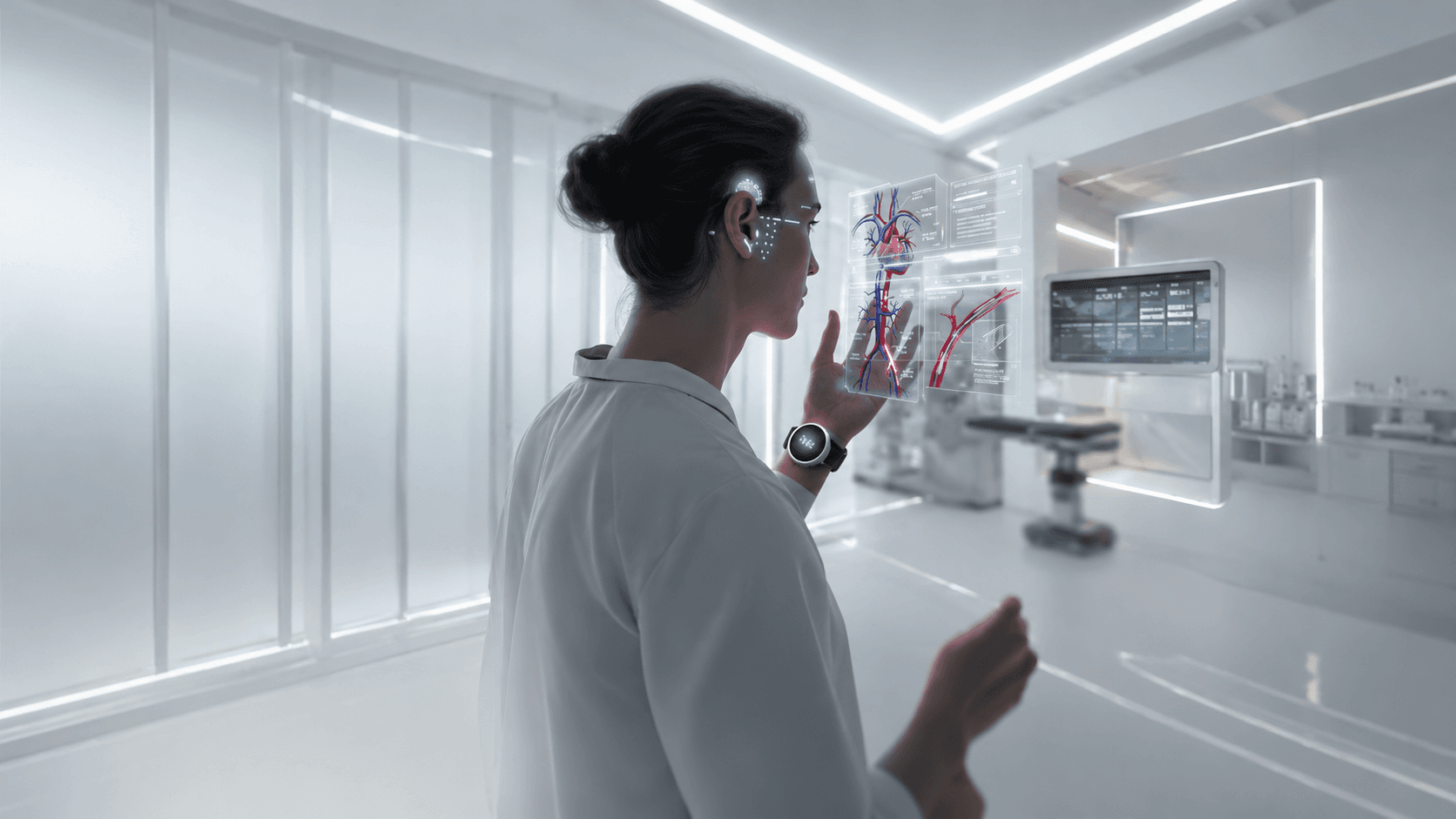
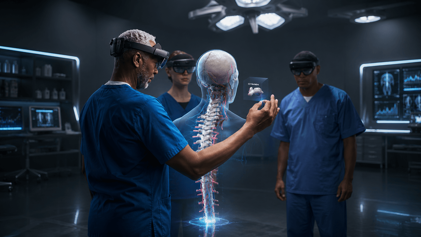
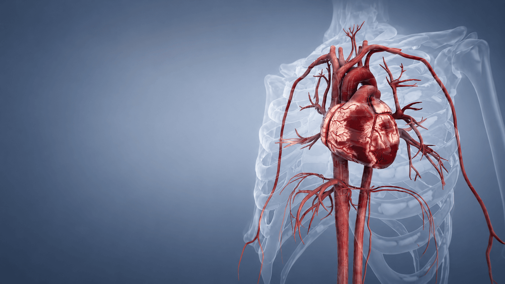
The core layer that turns CT, MRI, DICOM data, AI masks, and physician annotations into interactive spatial medical models.
  • Imaging and AI result ingestion
  • 2D slice and 3D model linkage
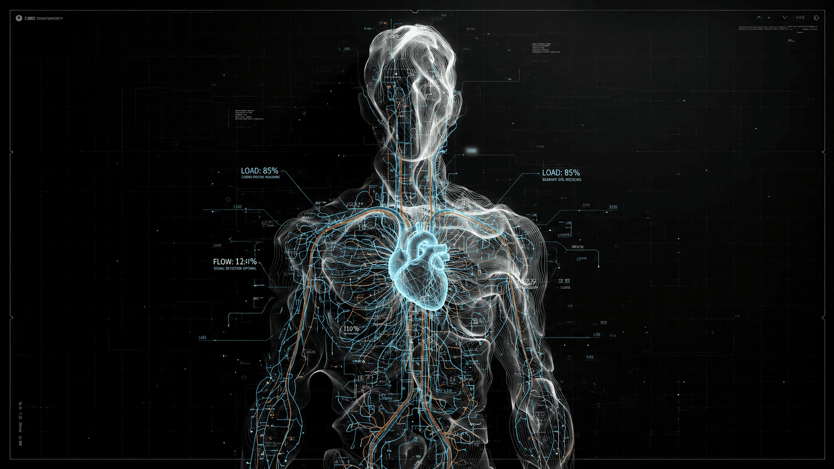
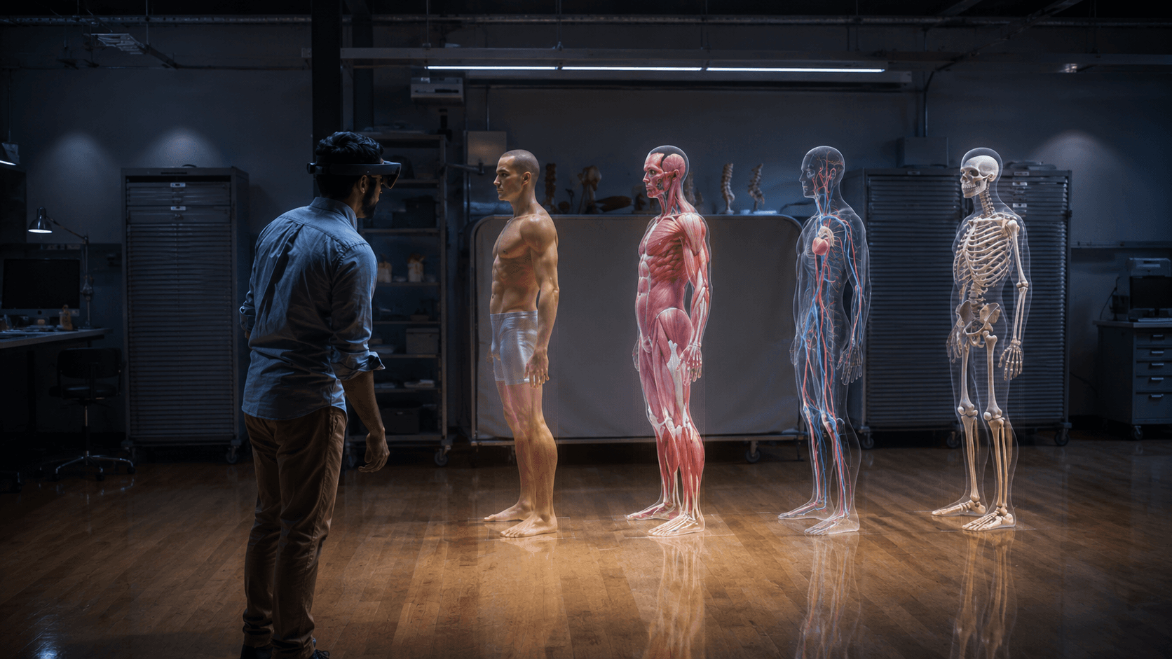
  • Lesion, organ, vessel, and bone layers

Medical schools, teaching departments, residency bases

Edu XR

Converts anonymized cases into layered teaching spaces for medical schools, hospital teaching departments, and residency training bases.
  • Case-based anatomy learning
  • Teacher-led annotations
  • Classroom, large-screen, and XR modes

Hospitals, specialty clinics, patient communication

Explain XR

Supports premium hospitals and specialty clinics with doctor-led spatial explanations that patients can understand.
  • Patient-friendly spatial views
  • Lesion and surrounding-structure context
  • Explanation snapshots or exports

MDT, radiology, remote expert collaboration

CaseRoom XR

Creates a shared case room for MDT teams, imaging departments, surgical teams, and remote experts to review one spatial model together.
  • Shared spatial case room
  • Synchronized markup and discussion
  • Remote expert participation

AI imaging, PACS/RIS, medical software partners

DeepView SDK

Gives AI imaging companies, PACS/RIS vendors, and medical software partners a spatial explanation layer for their outputs.
  • AI result ingestion
  • Embeddable 3D / XR viewer
  • Partner integration interface

Use Cases

Built around moments where spatial understanding changes the conversation.

Each scenario starts from an existing medical communication problem: teaching complex anatomy, explaining imaging to patients, aligning specialists, or making AI results reviewable.

01

Medical Education

Teaching rounds with layered real-case anatomy.

Medical schools, teaching departments, and residency bases can turn anonymized imaging cases into spatial lessons with teacher annotations and reusable case libraries.
02

Patient Communication

A clearer consultation around the image.

High-end hospitals and specialty clinics can help patients understand lesion location, boundaries, and nearby anatomy through doctor-led spatial explanation.
03

Case Discussion / MDT

One case space for multi-specialty review.

Radiology, surgery, oncology, and remote experts can review the same case model, mark regions together, and reduce context loss across meetings.
04

AI Result Explanation

AI outputs that doctors can verify in space.

Segmentation masks, lesion boundaries, and AI findings become spatial objects connected to anatomy, making review, teaching, and explanation easier.

Spatial Workflow

From pixels and masks to a shared medical space.

The workflow keeps physicians in control: source imaging and AI outputs become structured spatial content for education, explanation, review, and collaboration.

1Medical imaging data
2AI mask and physician annotation
3DeepView spatial reconstruction
4Education, explanation, review, collaboration

Technical Moat

Complex medical data, made spatial, interactive, collaborative, and deployable.

The technical story is not a headset demo. It is a visualization, interaction, collaboration, and cross-device foundation for medical imaging workflows.

High-resolution 3D visualization

Handles complex volumetric medical data with layered structure, depth, and spatial precision.

Medical volumetric interaction

Supports slicing, rotating, highlighting, measuring, and explaining structures in spatial context.

Immersive XR interaction

Adapts medical case views across PC, large displays, tablets, VR, and MR environments.

Multi-user collaboration

Gives physicians, educators, students, and remote experts a shared case space for discussion.

Cross-device deployment

Keeps the same case accessible without binding the platform to a single headset ecosystem.

AI result spatial explanation

Converts segmentation and lesion outputs into doctor-reviewable anatomy relationships.

Medical Boundary / Trust

Designed for responsible medical communication.

Pholoid is not a replacement for clinical diagnosis. It is designed for education, communication, review, collaboration, and doctor-led spatial explanation.

Not independent diagnosisNot intraoperative real-time navigationNot a treatment guarantee

Contact

Discuss a pilot for medical education, patient communication, case collaboration, or AI imaging partnership.

Bring one real workflow: a teaching case, a patient explanation moment, an MDT discussion, or an AI result that needs spatial interpretation.

International Collaboration

Regional Presence

Pholoid is building an international collaboration structure across Hanoi, Kuala Lumpur, and U.S.-based technical advisory resources for medical spatial intelligence.

01

Hanoi, Vietnam

Regional Headquarters & Market Development Base

Address: 25 Le Thanh Tong Street, Hanoi, Vietnam

Supporting Southeast Asia market development, institutional partnerships, government-facing collaboration, and regional business coordination.

02

Kuala Lumpur, Malaysia

Technology Hub & Product Engineering Center

Focused on DeepView XR Engine, medical imaging spatialization, AI segmentation visualization, XR interaction systems, and multi-device product engineering.

03

United States

Technical Advisory Network

U.S.-based technical advisory resources in immersive visualization, medical volumetric interaction, spatial computing, and high-resolution 3D display systems provide input for Pholoid’s product direction.